Introduction :
Growth and reproduction are characteristics of cells, indeed of all living
organisms. All cells reproduce by dividing into two, with each parent cell
giving rise to two daughter cells each time they divide. These newly formed
daughter cells can themselves grow and divide, giving rise to a new cell
population that is formed by the growth and division of a single parental
cell and its progeny. In other words, such cycles of growth and division
allow a single cell to form a structure consisting of millions of cells.
MEIOSIS
"Term meiosis" was proposed by Farmer and Moore.
The specialised kind of cell division that reduces the chromosome number by
half results in the production of haploid daughter cells. This kind of
division is called meiosis.
Meiosis ensures the production of haploid phase in the life cycle of sexually
reproducing organisms whereas fertilisation restores the diploid phase.
Meiosis occurs during gametogenesis, leads to the formation of haploid
gametes.
The key features of meiosis are as follows:
Meiosis involves two sequential cycles of nuclear and cell division called
meiosis I and meiosis II but only a single cycle of DNA
replication.
Meiosis I:-
Heterotypic division or reduction division. It leads to reduction in
chromosome numbers. Division of chromosome does not occurs in meiosis-1
land only segregation of homologous chromosomes takes place.
Meiosis-1 is initiated after the parental chromatids have replicated to
produce identical sister chromatids at the S-Phase.
Meiosis-1 involves pairing of homologous chromosomes and
recombination between non sister chromatids of homologous chromosome.
Meiosis II :
This is a homotypic division or equational division. It does not leads to
any change in chromosome number.
Division of chromosome or centromere occurs during meiosis II.
Four haploid cells are formed at the end of meiosis ll. All the
four daughter cells produced by meiosis are genetically different
from each other and also differ from the mother cell.
In meiosis, division of nucleus takes place twice but division of chromosome
occurs only once in meiosis-Il.
Meiotic events can be grouped under the following phases :
Meiosis I
Meiosis II
Prophase I
Prophase II
Metaphase I
Metaphase II
Anaphase I
Anaphase II
Telophase I
Telophase II
Interphase - same as in mitosis.
Stages of meiosis I
1. Prophase -1:
Typically longer and more complex when compared to prophase of mitosis.
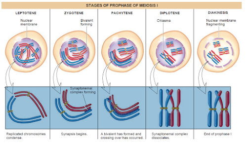
Prophase l is classified in five substages based on chromosomal
behaviour :
(a) Leptotene = Chromatin threads condense to form chromosomes.
Chromosomes are longest & thinest
Chromosomes become gradually visible under the light microscope.
All the chromosomes in nucleus remain directed towards centrioles, so group
of chromosomes in nucleus appears like a bouquet. (Bouquet stage).
(b) Zygotene or Synaptotene - Zygotene is characterized by
pairing of homologous. chromosomes (Synapsis). Pairs of homologous
chromosomes are called Bivalents or tetrads. However these are more
clearly visiblr at next stage (pachytene). A structure develops in
between homologous chromosomes. Which is termed as
synaptonemal complex.
The 1st two stages of prophase I is relatively short lived compared to
the pachytene.
( c) Pachytene (Thick thread) - Due to increased attraction,
homologous chromosomes tightly coil around each other. Both the chromatids
of each chromosome become distinct and are called sister chromatids.
During this stage, the four chromatids of each bivalent chromosome become
distinct and clearly appeared as tetrad.
Recombination nodules between nonsister chromatids of homologous pair
develop and these non sister chromatid exchange their parts. i.e. crossing over.
Crossing over leads to recombination of genetic material on the two
chromosomes.
Crossing over is an enzyme mediated process and the enzyme involved is
called recombinase (Endonuclease+ligase).
Recombination between homologous chromosomes is completed by the end of
pachytene, leaving the chromosomes linked at the sites of crossing over.
(d) Diplotene - The begining of diplotene is recognised by
dissolution of synaptonemal complex. Homologous chromosomes start repulsing
each other so X-shape structures.appeared called chiasmata.
Diplotene may last long up to months or years in oocytes of some vertebrates
(Dictyotene).
(e) Diakinesis - It is final stage of meiotic prophase I. Marked by
terminalization
of chiasmata (Chiasmata open in zip like manner).
Chromosome are fully condensed and meiotic spindle is assembled to prepare
the homologous chromosome for separation.
Centrioles move towards the opposite poles.
By the end of diakinesis nucleolus disappear and the nuclear envelope also
breaks down.
Diakinesis represents transition to metaphase.
2. Metaphase I:
Bivalents arrange on equator (congression) of cell to form metaphase
plate. The microtubules (spindle fibres) from the opposite poles of the
spindle attach to the pair of homologous chromosome with one kinetochore of
each chromosome.
Two types of spindle fibres appear in the cell:
(i) Chromosome / Kinetochore Spindle fibres
(ii) Supporting/Continuous /non-kinetochore Spindle fibres
3. Anaphase I:
Due to shortening of kinetochore/chromosomal fibres homologous chromosomes
segregate from each other and move towards the opposite poles. Sister
chromatids remain associated at their centromeres (i.e. chromosomes remain
in double chromatid stage).
Anaphase I is characterised by segregation or disjunction of
chromosomes. Division of centromere is absent.
4. Telophase I :
The nuclear membrane and nucleolus reappear. Although in many case the
chromosomes do undergo some dispersion, but they do not reach the extremely
extended state of the interphase nucleus.
Cytokinesis follows telophase-l and a diploid (2n) cell divides into two
haploid (n) daugther cells. This is called as dyad of cells.
Interkinesis :- Gap between meiosis l and meiosis II is called
Interkinesis. Preparations of meiosis ll occur during interkinesis.
It is like interphase of mitosis but
replication of DNA is absent in interkinesis.
Interkinesis is generally short lived. Interkinesis is followed by
prophase-Il, a much simpler prophase than prophase-l.
Stages of Meiosis - II
1. Prophase II :
Meiosis ll dis initiated immediately after cytokinesis, usually before the
chromosomes have fully elongated. In contrast to meiosis l, meiosis ll
resembles a normal mitosis. The nuclear membrane disappears by the end of
prophase II. The chromosomes again become compact.
2. Metaphase II
At this stage the chromosomes align at the equator and the microtubules from
opposite poles of the spindle get attached to the kinetochores of sister
chromatids.
3. Anaphase II:
It begins with the simultaneous splitting of the centromere of each
chromosome (which was holding the sister chromatids together), allowing them
to move toward opposite poles of the cell by shortening of microtubules
attached to kinetochores.
4. Telophase II:
Meiosis ends with telophase II, in which the two groups of chromosomes once
again get enclosed by a nuclear envelope; cytokinesis follows resulting in
the formation of tetrad of cells i.e. four haploid daughter
cells.
Significance of Meiosis :
(1) Meiosis is the mechanism by which
conservation of specific chromosome number of each species is
achieved across generations in sexually reproducing organisms, even though
the process (per se paradoxically) results in reduction of chromosome number
by half.
(2) It also increases the genetic variability in the population of
organisms from one generation to the next. Variations are very important for
the process of evolution.
NOTE : Prophase-I further subdivide into five phases based on
the chromosomes behaviour.
- Meiosis ensures the production of haploid phase in the life cycle of sexually reproducing organism.
- Meiosis involves pairing of homologous chromosomes and recombination between them.
- Chiasmata formation is the result of crossing over.
- Meiosis increases the genetic variability in the population of organism from one generation to next.


0 comments:
Post a Comment
If you have any doubts. Please let me know.