Wednesday, July 8, 2020

DNA REPLICATION

RNA WORLD

RNA was the first genetic material. There are evidence to suggest that essential life processes, such as metabolism, translation, splicing etc. evolved around RNA. RNA use to act as a genetic material as well as a catalyst, there are some important biochemical reactions in living system that are catalysed by RNA catalysts and not by Protein enzymes (e.g. splicing) RNA being a catalyst was reactive and hence unstable. Therefore, DNA has evolved from RNA with chemical modification that makes it more stable. DNA being double stranded and having complementary strand further resists changes by evolving a process of repair. RNA is adapter, structural molecule and in some case catalytic. Thus RNA is better material for transmission of information. 


DNA REPLICATION


1. While proposing the double helical structure for DNA, Watson and Crick had immediately proposed a scheme for replication of DNA. To quote their original statement that is as follows.

2. "It has not escaped our notice that the specific pairing we have postulated immediately suggests a possible copying mechanism for the genetic material"           ( Watson and Crick, 1953).
 
3. The scheme suggested that the two strands would separte and act as a template for the synthesis of new complementary strands. After the completion of replication, each DNA molecule would have one parental and one newly synthesised strand. This scheme was termed as semiconservative DNA replication.

4. DNA capable of self duplication.

5. All living beings have the capacity to reproduce because of this characteristic of DNA.

6. DNA replication takes place in "S-phase" of the cell cycle. At the time of cell division, it divides in equal parts in the daughter cells.

SEMI CONSERVATIVE MODE OF DNA REPLICATION


Semi conservation mode of DNA replication was first proposed by Watson and Crick. Later on it was experimentally proved by Meselson and Stahl (1958) in E.coli and Taylor in Vicia faba (1958). To prove this method, Taylor used Radiotracer Technique in which Radioisotopes (tritiated thymidine = ) were used. Meselon and Stahl used heavy isotope of nitrogen-15.


Matthew Meselson and Franklin Stahl performed the following experiment in 1958:

1. They grew E.coli in a medium containing NH4CL ( N-15 is the heavy isotope of nitrogen) as the only nitrogen source for many generations. The result was that N-15 was incorporated into newly synthesised DNA (as well as other nitrogen containing compounds). This heavy DNA molecule could be distinguished form the normal DNA by centrifugation in as cesium chloride (CsCl) density gradient (please note that N-15 is not a radioactive isotope, and it can be separated from N-14 only based on densities).

2. Then they transferred the cells into a medium with normal NH4CL and took samples at various definite time intervals as the cells multiplied, and extracted the DNA that remained as double stranded helices. The various samples were separated independently on CsCl gradient to measure the densities of DNA.

3. Thus, the DNA that was extracted from the culture one generation after the transfer from N-15 to N-14 medium [that is after 20 minutes; E.coli divides in 20 minutes] had a hybrid or intermediate density. DNA extracted from the culture after another generation [that is after 40 minutes, 2 generation] was composed of equal amounts of this hybrid DNA and of 'light' DNA.


MECHANISM OF DNA REPLICATION
 The following steps are included in DNA replication:-
1. Unzipping (Unwinding):-
 
1. The separation of 2 chains of DNA is termed as unzipping. And it takes place due to the breaking of H-bond. The process of unzipping starts at a certain specific points which is termed as initiation point or origin of replication. In prokaryotes there occurs only one origin of replication but in eukaryotes there occur many origin or replication i.e unzipping starts at many points simultaneously. At the place of origin the enzyme responsible for unzipping (breaking the hydrogen bond) is Helicase (=Swivelase). In the process of unzipping Mg+2 act as cofactor.

2. SSB (Single stranded DNA binding protein) prevents the information of H-bonds.

3. Topoisomerase (in prokaryotes also called as DNA gyrase)  release the tension arises due to supercoiling. 
Note: The process of DNA replication takes a few minutes in prokaryotes and a few hours in Eukaryotes.


2. Formation of New chain:- 
 
1. To start the synthesis of new chain, special type of RNA is required which is termed as RNA primer. The formation of RNA primer is catalysed by an enzyme- RNA Polymerase (primase). Synthesis of RNA-Primer takes place in 5'-3' direction. After the formation of New chain, this RNA is removed. For the information of new chain Nucleotides are obtained from Nucleoplasm. In the Nucleoplasm, Nucleotides are present in the form of triphosphates like dATP, dGTP, dCTP, dTTP etc. 

2. During replication, the 2 phosphate groups of all nucleotides are separated. In this process energy is yeilded which is consumed in DNA replication.

3. Energetically replication is a very expensive process. Deoxyribonucleoside triphosphase serve dual purposes in addition to acting as substrates they provide energy for Polymerisation. 


4. The formation of New chain always takes place in 5'-3' direction. As a result of this, one chain of DNA is continuously formed and it is termed as Leading strand. The formation of second chain begins from the centre and not form the terminal points, so this chain is discontinuous and is made up of small segments called Okazaki fragments. This discontinuous chain is termed as Lagging strand. Ultimately all these segments joined together and a complete new chain if formed. 

5. The Okazaki fragments are joined together by an enzyme DNA Ligase.

6. The formation of New chains in catalysed by an enzyme DNA Polymerase. In prokaryotes it is of 3 types:

1. DNA-polymerase 1 :- This was discovered by KORNBERG (1957). So it is also called as 'Kornberg's enzyme'. KORNBERG also synthesized DNA first of all, in the laboratory. This enzyme functions as exonuclease. It separates RNA-Primer from DNA and also fills the gap. It is also known as DNA-repair enzyme.

2. DNA-polymerase 2 :-  It is least reactive in replication process. It is also helpful in DNA-repairing in absence of DNA-polymerase 1 and DNA-polymerase 3.

3. DNA-polymerase 3 :- This is the main enzyme in DNA-replication. It is most important. The larger chains are formed by this enzyme. This is also known as Replicase. 

7. In the semi conservative mode of replication each daughter DNA molecule receives one chain of polynucleotides from the mother DNA- molecule and the second chain is synthesized.


Sepcial points:-
1. All DNA polymerase 1,2 and 3 enzymes have 5'-3' Polymerisation activity and 3'-5' exonuclease activity.

2. DNA polymerase 1 also has 5'-3' exonuclease activity.

3. Any failure in cell division after DNA replication result into polyploidy.

4. Difference between DNAs and DNase is that DNAs means many DNA and DNase means DNA digestive enzymes.


Thank you👍

Tuesday, July 7, 2020

Molecular Basis of Inheritance NUCLEIC ACIDS

NUCLEIC ACIDS

1. F. Meischer discovered nucleic acid in nucleus of pus cell and called it "nuclein". The term nucleic acid was coined by "Altman".

2. Nucleic acids are polymer of nucleotides.
Example: DNA and RNA.

Nucleotide= Nitrogen base + Pentose sugar + phosphate.

Nucleoside= Nitrogen base + Pentose sugar.

A. Nitrogen base: 
On the basis of structure nitrogen bases are broadly of two types:- 
1. Pyrimidines- consist of one pyrimidine ring. Skeleton of ring composed of two nitrogen and four carbon atoms. e.g. Cytosine, Thymine and  Uracil.

 2. Purines- consist of two rings i.e. one pyrimidine ring (2N+4C)  and one imidazole ring (2N+3C) e.g. Adenine and Guanine.

B. Pentose sugar (Number of carbon= 5):-
C. Phosphate:
-Acidic
-Negative charged

Nitrogen base froms bond with first carbon of pentose sugar to form a nucleoside. Nitrogen of first place (N1) forms bond with sugar in case of pyrimidines while in purines nitrogen of ninth place (N9) forms bond with sugar.

Phosphate forms ester bond (covalent bond) with fifth carbon of sugar to form a complete nucleotide.


Types of Nucleosides and Nucleotides

1. Adenine+Ribose=Adenosine
Adenosine+Phosphate=Adenylic acid (AMP).

2. Adenine+Deoxyribose=Deoxy adenosine
Deoxyadenosine+P=Deoxyadenylic acid (dAMP).

3. Guanine+Ribose=Guanosine
Guanosine+P=Cytidylic acid (CMP).

4.Guanine+Deoxyribose=Deoxy Guanosine
Deoxy Guanosine+P= Deoxy guanylic acid (dGMP).

5. Cytosine+Ribose=cytidin
Cytidine+P=Cytidylic acid (CMP).

6. Cytosine+Deoxyribose=Deoxycytidine
Deoxycytidine+P=Deoxycytidylic acid (dCMP).

7. Uracil+Ribose=Uridine 
Uridine+P=Uridylic acid (UMP).

8.Thymine+Deoxyribose=Deoxy thymidine
Deoxycytidine+P= Deoxythymidylic acid (stamp).

DNA


1.DNA as an acidic substance present in nucleus was first identified by Friedrich Meischer in 1869.

2. DNA term was given by Zacharis.

3. DNA is long polymer of deoxyribonucleotides.

4. DNA is negatively charged.

5. In DNA pentose sugar is Deoxyribose sugar and for types of nitrogen bases A,T,G,C.

6. Wilkins and Franklin studied DNA molecule with help of X-Ray cyrstallography.

7. With the help of this study, Watson and Crick (1953) proposed a double helix model for DNA. For this model Watson, Crick and Wilkins were awarded by Nobel Prize in 1962.

8. One main hallmark (main point) of double helix model is complementary base pairing between purine and pyrimidine.

9. According to this model, DNA is composed of two polynucleotide chains.

10. Both polynucleotide chains are complementary and antiparallel to each other.

11. In both strand of DNA direction of phosphodiester bond is opposite. i.e. If direction of phosphodiester bond in one strand is 3'-5' then it is 5'-3' in another strand.

12. Both strand of DNA are held together by hydrogen bonds. These hydrogen bonds are present between nitrogen bases of  both  strand. 

13. Adenine binds to thymine by two hydrogen bonds and Cytosine binds to guanine by three hydrogen bonds. 

14. In a DNA molecule one purine always pairs with pyrimidine. This generates approximately uniform distance between the two strand of DNA.

15. In DNA plane of one bases pair stacks over the other in double helix. This, in addition to H-bonds, confers stability of the helical structure of DNA.

16. Chargaff's equivalency rule- In double stranded DNA amount of purine nucleotides is equals to amount of pyrimidine Nucleotides.

Purine=Pyrimidine
 [A]+[G]
------------ =1
 [T]+[C] 

17. Base ratio:- 
 A+T
------ = constant for given species. i.e         
G+C                 species specific.

18. In DNA A+T>G+C = A-T type DNA. Base ratio of A - T type of DNA is more than one. Eg. Eukaryotic DNA.

19. In a DNA G+C>A+T= G-V type DNA. Base ratio of G - C type of DNA is less than one.
Eg. Prokaryotic DNA.

20. Melting point of DNA depends on G-C contents.

More G-C contents means higher melting point.
Tm= Temperature of melting.
Tm of prokaryotic DNA>Tm of Eukaryotic DNA.

21. DNA absorbs U.V. rays of 2600A° wavelength.

22. Denaturation and renaturation of DNA - If a normal DNA molecule is placed at high temperature (80-90°c) then both strands of DNA will separate from eachother due to breaking of hydrogen bonds. It is called DNA-denaturation. 


23. When denatured DNA molecule is placed at normal temperature then both strand of DNA attached and recoiled to each other. It is called renaturation of DNA.


Configuration of DNA molecules:-

1. Two strands of DNA are helically coiled like a revolving ladder. Backbone of this ladder (Reiling) is composed of phosphates and sugars while steps (bars) are composed of pairs of nitrogen bases.
Two chains have anti-parallel polarity. It means, if one chain has the polarity 5'-3', the other has 3'-5'.


2. Distance between two successive steps is 3.4A°. In one complete turn of DNA molecule there are such 10 steps (10 pairs of nitrogen bases). So the length of one complete turn is 34A°. This is called helix length.

3. Diameter of DNA molecule i.e. distance between phosphates of two strands is 20A°.

4. Each step of ascent is represented by a pair of bases. At each step of ascent, the strand turns 36°.

5. In nucleus of Eukaryotes the DNA is associated with histone protein to form nucleoprotein.

6. Bond between DNA and Histone is salt linkage (Mg+2).

ϕ ×174 (bacteriophage) [single stranded] - 5386 Nucleotides.
λ bacteriophage - 48502 base pair.
E. Coli - 4.6× 106  base pair.
Human - 6.6× 109  base pair.

Types of DNA:-
On the basis of direction of twisting, there are two types of DNA.
1. Right Handed DNA- clockwise twisting e.g. the DNA for which Watson and Crick proposed model was 'B'DNA.
2. Left Handed DNA- Anticlockwise twisting e.g. Z-DNA - Discovered by Rich. Phosphate and sugar backbone is zig-zag.

Helix length - 45.6 A°.
Diameter - 18.4 A°.
No. Of base pairs - 12 (6 dimers).
Distance between 2 bases pairs - 3.75 A°.

Palindromic DNA- Wilson and Thomas.
 
CC   GG  TA  CC  GG
GG   CC  AT  GG  CC 
                                 ← 
Sequence of nucleotides same form both ends.


PACKAGING OF DNA HELIX


The average distance between the two adjacent base pairs of 0.34 nm (0.34 x 109 m or 3.4 A°). Length of DNA for human diploid cell is 6.6.x 109 bp x 0.34 x 109 m/bp =2.2m. The length is far greater than the dimension of typical nucleus (approximately 10–6 m).  The number of bade pairs in Escherichia coli is 4.6×10⁶ . The total length is 1.36 mm. The long sized DNA accommodated in small area (about 1  μm in  E. coli) only through packing of compacted. DNA s acidic due to presence of large number of phosphate group. Compaction occurs by folding acid attachment of DNA with basic proteins, polyamine in prokaryotes and histone in eukaryotes.

DNA packaging in Prokaryotes : DNA is found in cytoplasm in supercoiled state. The coils and maintained by non histone basic protein like polyamines. This compact structure of DNA is called nucleoid genophore.

DNA packaging in Eukaryotes: It is carried out with the help of lysine and arginine rich basic proteins called histone. The unit of compaction is nucleosome. There are five types of histone proteins H1,H2A,H2B,H3 and H4.  

Four of them occur in pairs to produce histone octamer (2 copies of each- H2A,H2B,H3 and H4), called nubody or  core of nucleosome. Their positively charged ends are directed outside. The negatively charged DNA is wrapped around the positively charged histone octamer to form a structure called nucleosome. A typical nucleosome contains 200 bp of DNA Helix. DNA present between two adjacent nucleosome is called linker DNA. It is attached to H1 histone protein. Length of linker DNA varies from species to species. Nucleosome chain gives a beads on string appearance under electron microscope. The nucleosomes furthers coils to form solenoid. It has diameter  of 30 nm as found in chromatin The beads on string structure in chromatin is packaged to  form chromatin fibres that are further coiled and condensed at metaphase stage of cell division to form chromosomes. The packaging at higher level requires additional set of proteins (acidic) that collectively are referred to as non histone chromosomal (NHC) Proteins.

Non-Histone chromosomal proteins are of three types:

1.Structural NHC protein.
2.Functional NHC protein e.g. DNA polymerase, RNA polymerase.
3.Regulatory NHC protein

In a typical nucleus, some region of chromatin are loosely packed (and stains light) and are referred to as euchromatin. The chromatin that is more densely packed and stains dark is called as heterochromatin, specifically euchromatin is said to be transcriptionally active and heterochromatin is transcriptionally inactive.


Thank you😋


LEAVE A COMMENT BELOW..................

Monday, July 6, 2020

CELL CYCLE AND CELL DIVISION (MITOSIS)

Introduction :

Growth and reproduction are characteristics of cells, indeed of all living organisms. All cells reproduce by dividing into two, with each parent cell giving rise to two daughter cells each time they divide. These newly formed daughter cells can themselves grow and divide, giving rise to a new cell population that is formed by the growth and division of a single parental cell and its progeny. In other words, such cycles of growth and division allow a single cell to form a structure consisting of millions of cells.


Cell division is of two types :


MITOSIS

Term mitosis was proposed by Flemming. Mitosis produces genetically identical cells, which are similar to mother cell.


Cause of mitosis :

(i) Kern plasm theory: Hertwig proposed kern plasm theory. According to this theory mitosis occurs due to disturbance in Karyoplasmic Index (KI) or Nucleocytoplasmic ratio of cell.

Karyoplasmic Index: 
  • Karyoplasmic Index of small cell is high as they have less cytoplasm. Nucleus efficiently controls the activity of cytoplasm in small cells.
  • In a large cell nucleus fail to control the activity of cytoplasm. To attain the control of nucleus on metabolism a large cell divides into two cells.

(ii) Surface-volume Ratio:
  • Surface volume ratio is also considered as a cause of cell division. When a cell grows in size its volumes increases more than its surface. So a stage will reach when the surface area becomes insufficient to draw the material. At such critical stage, division of cell started.

CELL CYCLE

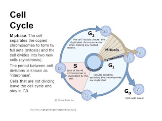
  • Cell division is a very important process in all living organisms. During the division of a cell, DNA replication and cell growth also take place.
  • All these processes, i.e.cell division, DNA replication, and cell growth. hence, have to take place in a coordinated way to ensure correct division and formation of progeny cells containing intact genomes.
  • The sequence of events by which a cell duplicates its genome, synthesises the other constituents of the cell and eventually divides into two daughter cells is termed cell cycle.
  • Although cell growth (in terms of cytoplasmic increase) is a continuous process.DNA synthesis occurs only during one specific stage in the cell cycle.
  • The replicated chromosomes (DNA) are then distributed to daughter nuclei by a complex series of events during cell division. These events are themselves under genetic control.

PHASES OF CELL CYCLE

  • A typical eukaryotic cell cycle is illustrated by human cells in culture. These cells divide once in approximately every 24 hours.
  • Yeast can progress through the cell cycle in only about 90 minutes. The time period of cell cycle is varied from organism to organism and also from cell type to cell type.

Cell cycle involves two stages -
(1) Interphase 
(2) Division phase/M-phase

1. Interphase :- This is phase between two successive M-phase. In interphase cell grows in size and prepares itself for next division. Interphase is most active phase of cell cycle. The interphase last more than 95% of the duration of cell cycle.
  • A series of metabolic changes occurs during interphase in cell. These changes were not visible under microscope, So some scientist termed interphase as resting phase. It is the time during which cell is preparing for division by undergoing both cell growth and DNA replication in an orderly manner.

Howard and Pelc classified interphase into three sub stages:- 

(i) G1-phase or Pre DNA synthesis phase (1st Gap phase)
  • G1 phase corresponds to the interval between mitosis and initiation of DNA replication. During G1, phase the cell is metabolically active and continuous grows.
  • During G1, most of cell organelles increases in cell and cell rapidly synthesizes different types of RNA and proteins. Due to availability of protein, synthesis of new protoplasm takes place in cell ard it starts growing in size. Cell grows maximum in G1 stage.

(ii) S- phase (DNA synthesis phase):

  • Replication of nuclear DNA and synthesis of histone protein takes place in s-phase. Replication of cytoplasmic DNA may occur in any stage of cell cycle.
  • During this time the amount of DNA per cell doubles. If the  Initial Amount of DNA is denoted as 2C then It increases to 4C. However, there is no increase in the chromosome number: if the cell has diploid or 2n number of chromosomes at G1, even after S-Phase the number of chromosomes remains the same, i.e., 2n.
  • S-Phase marks the phase of DNA replication and chososome duplication (DNA content in chromosome become double).
  • In animal cells, during the S-phase, DNA replication begins in the nucleus, and the centriole duplicates in the cytoplasm.

G2- phase (2nd Gap phase) or Post DNA synthesis phase (Pre mitosis phase)

Actual preparation (Final preparation) of M-phase occurs during this phase. Special materials required for M-phase are synthesized in G1 phase. eg. Tubulin protein. -(Required for formation of spindle fibres). Cell growth continues.

G0 phase-
  •  Some cells in the adult animals do not appear to exhibit division (e.g. heart cells) and many other cells divide only occasionally, as needed to replace cells that have been lost because of injury or cell death.These cells that do not divide further exit G1 phase to enter an inactive  stage called quiescent stage (G0) of the cell cycle.
  • Cells in this stage remain metabolically active but no longer proliferate (divided) unless called on to do so depending on the requirement of the organism.


Checkpoints of cell cycle :

  • Cell cycle is running by a group of special proteins "Cyclins and Cdks (MPF). (Nurse, T.Hunt & Hartwell 2001 studies on saccharomyces)
  • Cell cycle is running by a group of special proteins "Cyclins and Cdks.
  • The activity of enzymes, known as cyclin dependant kinases. (Cdk's) regulates the cell cycle. Kinase is an enzyme that removes a phosphate group from ATP & add to another protein. The kinases involved in the cell cycle are called Cdks because they are activated when they combined with key protein called cyclin.
  • At some check points a kinase enzyme combines with cyclin & this moves the cell cycle forwardly.
  • G2-M transition is triggered by maturation promoting factor (MPF) formed by M-cyclin + CDK2.


2. Division phase :

Division phase or M-phase or mitotic phase lasts for only about an hour in the 24 hour duration of cell cycle of a human cell.


The M-phase represents the phase when the actual cell division or mitosis occurs.

In animals, mitotic cell division is restricted or only seen in diploid somatic cell except in some social insects. Against this, the plants can show mitotic division in both haploid and diploid cells.

This is the most dramatic period of the cell cycle, involving a major reorganisation of virtually all components of the cell. Since the number of chromosomes in the parent and progeny cells is the same, it is also called as equational division.

Though for convenience mitosis has been divided into four stages of nuclear division, it is very essential understand that cell division is a progressive process and very clear-cut cannot be drawn between various stages.

The M-phase start with nuclear division, corresponding to the separation of daughter chromosome (Karyokinesis) and usually ends with division of cytoplasm (cytokinesis).

Mitosis is divided into the following four stages :
  *Prophase *Metaphase *Anaphase *Telophase 

1. Prophase :

Prophase which is the first stage of karyokinesis of mitosis follows the S And G2 phases of interphase.

In the S and G2 phase the new DNA molecules formed are not distinct but Intertwined.

Prophase is marked by the initiation of condensation of chromosomal material. The chromosomal material becomes untangled during the process of chromatin condensation.

The centriole, which had undergone duplication during S-phase of interphase, now begins to move towards opposite poles of the cell.

Formation of astral ray occurs due to gelation of proteins around centrioles in animal cells.

Anastral and Amphiastral Mitosis: In higher plants, centrioles are absent and no asters are formed. Mitosis without asters is known as anastral mitosis. In animals, the asters are present and the mitosis is described as amphiastral or astral mitosis.

The completion of prophase can thus be marked by the following characteristic events:
  •  Chromosomal material condenses to form compact mitotic chromosomes. Chromosomes are seen to be composed of two chromatids attached together at the centromere.
  • Centrosome which had undergone duplication during interphase, begins to move towards opposite poles of the cell. Each centrosome radiates out microtubules called asters. The two asters together with spindle fibres forms mitotic apparatus.
  • Cell at the end of prophas when viewed under the microscope, do not show golgi complexes, endoplasmic reticulum, nucleolus and nuclear envelope.

(2) Metaphase

The complete disintegration of the nuclear envelope marks the start of the second phase of mitosis, hence the chromosomes are spread through the cytoplasm of the cell.

By this stage, condensation of chromosomes is completed and they can be observed clearly under the microscope.This then, is the stage at which morphology of chromosomes is most easily studied.

At This stage, metaphase chromosome is made up of two sister chromatids, which are held together by the centromere. Small disc-shaped structures at the surface of the centromeres are called kinetochores. These structures serve as the sites of attachment of spindle fibres (formed by the microtubules) to the chromosomes that are moved into position at the centre of the cell.


Hence, the metaphase is characterised by all the chromosomes coming to lie at the equator with one chromatid of each chromosome connected by its kinetochore to spindle fibres from one pole and its sister chromatid connected by its kinetochore to spindle fibres from the opposite pole. The plane of alignment of the chromosomes at metaphase is referred to as the metaphase plate.

Chromosomal fibres (discontinous/kinetochore which run from pole to centromere) and supporting fibres (continous/non-kinetochore, which run from pole to pole) arrange in cell.

Centromere lies at equator and arms of chromosomes remain directed towards poles.

The key features of metaphase are:
  • Spindle fibres attach to kinetochores of chromosomes.
  • *Chromosomes are moved to spindle equator and get aligned along metaphase plate through spindle fibres to both poles.

(3) Anaphase

Centromere of each chromosome splits simultaneously lengthwise (division of centromere). Sister chromatids separate from each other and separated each chromatid is now reffered to as individual chromosome.

Number of chromosome become double in cell.

As each chromosome moves away from the equatorial plate, the centromere of each chromosome is towards the pole and hence at the leading edge, with the arms of the chromosome trailing behind.

The two new daughter chromosomes begin moving toward opposite ends of the cell as their kinetochore microtubule shorten due to depolymerisation of tubulin protein towards kinetochoric end. Because these microtubules are attached at the centromere region, the centromeres are pulled ahead of the arms. (Pulling).

The cell elongates as the nonkinetochore microtubules lengthen.
  • Anaphase stage is characterised by the following key events:
  1. Centromeres split and chromatids separate.
  2. Chromatids (now referred as chromosomes) move to opposite poles.

(4) Telophase (Reverse prophase):

 At the beginning of the final stage of karyokines, i.e.  telophase, the chromosomes that have reached their respective poles decondense and lose their individuality. The Individual chomosomes can no longer be seen and chromatin material tends to collect at each of the two poles. This in the stage which shows the following key events:

  • Chromosomes cluster at opposite spindle poles and their identity is lost as discrete elements.
  • Nuclear envelope develops around the chromosome dusters at each pole forming  two daughter nuclei.
  • Nucleolus, golgi complex and ER reform.


CYTOKINESIS

Mitosis accomplishes not only the segregation of duplicated chromosome into daughter nuclei (Karyokinesis) but the cell itself is divided into two daughter cells by the separation of cytoplasm called cytokinesis at the end of which cell division gets completed.

In animals cytokinesis occurs by constriction & furrow formation. Microtubules and microfilaments arrange on equator to form midbody and at the periphery of the equator a contractile ring is formed that is made up of actin and myosin protein. 
Due to interaction between actin and myosin ring contract, thus a furrow forms from outside to inside in cell. Furrow deepens continuously and ultimately a cell divides into two daughter cells. In animals cytokinesis occurs in centripetal order.


Cytokinesis in plants takes place by cell plate formation because constriction is not possible due to presence of the rigid cell wall. Many golgi vesicles and spindle microtubules arrange themselves on equator to form phragmoplast. Fragmentes of ER may also deposit in phragmoplast. Membrane of golgi vesicles fuse to for a plate like structure called cell plate. Golgi vesicles secrete calcium and magnesium pectate. Further cell plate is modified into middle lamella. In plants, cytokinesis occurs in centrifugal order (cell plate formation is from center to periphery).

In some organisms karyokinesis is not followed by cytokinesis as a result of which multinucleate condition arises leading to the formation of syncytium (e.g. liquid endosperm in coconut).

SIGNIFICANCE OF MITOSIS

Development of an organism occurs by mitosis. Every organism starts its life from a single cell i.e. zygote. Repeated mitosis in zygote leads to the formation of the whole body.

The growth of multicellular organisms is due to mitosis.

Cell growth results in disturbing the ratio between the nucleus and the cytoplasm. It therefore becomes essential for the cell to divide to restore the nucleo-cytoplasmic ratio.

A very significant contribution of mitosis is cell repair. The cells of the upper layer of the epidermis, cells of the living of the gut, and blood cells are being constantly replaced.

Mitotic divisions in the meristematic tissues - the apical (primary meristem) and the lateral cambium (secondary meristem), result in a continue growth of plants throughout their life.

MODIFICATIONS OF MITOSIS

Free nuclear division :- Karyokinesis is not followed by cytokinesis as a result of which multinucleated condition arises.

Endomitosis :- This is duplication of chromosomes without division of nucleus. Endomitosis leads to polyploidy.
i.e. Increase in number of set of chromosomes. Colchicine induces polyploidy in plants. Colchicine is a mitotic poison as it arrests the formation of spindle fibres

Endoreduplication :- Endoreduplication is a modification of endomitosis. The polytene chromosomes are formed by the process of endoreduplication. In endoreduplication, the chromatids replicate but do not get seperated. This process is also known as polyteny.

Note :

AMITOSIS : It is a simple method of cell division which is also called direct cell division. In this division there is no differentiation of chromosomes and spindle. The nuclear envelope does not degenerate. The nucleus elongates and constricts in the middle to form two daughter nuclei. This is followed by a centripetal constriction of the cytoplasm to form two daughter cells, eg. Prokaryotes and Some unicellular eukaryotes.


CELL CYCLE AND CELL DIVISION (MEIOSIS)

Introduction :

Growth and reproduction are characteristics of cells, indeed of all living organisms. All cells reproduce by dividing into two, with each parent cell giving rise to two daughter cells each time they divide. These newly formed daughter cells can themselves grow and divide, giving rise to a new cell population that is formed by the growth and division of a single parental cell and its progeny. In other words, such cycles of growth and division allow a single cell to form a structure consisting of millions of cells.


Cell division is of two types :


MEIOSIS

"Term meiosis" was proposed by Farmer and Moore.

The specialised kind of cell division that reduces the chromosome number by half results in the production of haploid daughter cells. This kind of division is called meiosis.

Meiosis ensures the production of haploid phase in the life cycle of sexually reproducing organisms whereas fertilisation restores the diploid phase. Meiosis occurs during gametogenesis, leads to the formation of haploid gametes.

The key features of meiosis are as follows:

Meiosis involves two sequential cycles of nuclear and cell division called meiosis I and meiosis II but only a single cycle of DNA replication.

Meiosis I:-

Heterotypic division or reduction division. It leads to reduction in chromosome numbers. Division of chromosome does not occurs in meiosis-1 land only segregation of homologous chromosomes takes place.
Meiosis-1 is initiated after the parental chromatids have replicated to produce identical sister chromatids at the S-Phase.

Meiosis-1 involves pairing of homologous chromosomes and recombination between non sister chromatids of homologous chromosome.

Meiosis II :

This is a homotypic division or equational division. It does not leads to any change in chromosome number.
Division of chromosome or centromere occurs during meiosis II. 
Four haploid cells are formed at the end of meiosis ll. All the four daughter cells produced by meiosis are genetically different from each other and also differ from the mother cell.

In meiosis, division of nucleus takes place twice but division of chromosome occurs only once in meiosis-Il.

Meiotic events can be grouped under the following phases :

Meiosis I                         Meiosis II 
      
Prophase I                       Prophase II
Metaphase I                    Metaphase II
Anaphase I                      Anaphase II
Telophase I                     Telophase  II

Interphase - same as in mitosis.

 Stages of meiosis I

1. Prophase -1:

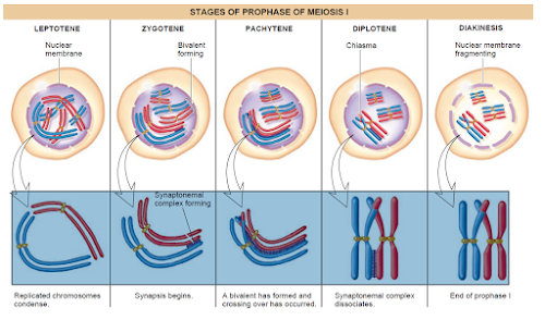
Typically longer and more complex when compared to prophase of mitosis. Prophase l  is classified in five substages based on chromosomal behaviour :


(a) Leptotene = Chromatin threads condense to form chromosomes. Chromosomes are longest & thinest
Chromosomes become gradually visible under the light microscope.

All the chromosomes in nucleus remain directed towards centrioles, so group of chromosomes in nucleus appears like a bouquet. (Bouquet stage).

(b)  Zygotene or Synaptotene - Zygotene is characterized by pairing of homologous. chromosomes (Synapsis). Pairs of homologous chromosomes are called Bivalents or tetrads. However these are more clearly visiblr at  next stage (pachytene). A structure develops in between homologous chromosomes. Which is termed as synaptonemal complex.

 The 1st two stages of prophase I is relatively short lived compared to the pachytene.

( c) Pachytene (Thick thread) - Due to increased attraction, homologous chromosomes tightly coil around each other. Both the chromatids of each chromosome become distinct and are called sister chromatids.

During this stage, the four chromatids of each bivalent chromosome become distinct and clearly appeared as tetrad.

Recombination nodules between nonsister chromatids of homologous pair develop and these non sister chromatid exchange their parts. i.e. crossing over.

Crossing over leads to recombination of genetic material on the two chromosomes.

Crossing over is an enzyme mediated process and the enzyme involved is called recombinase (Endonuclease+ligase).

Recombination between homologous chromosomes is completed by the end of pachytene, leaving the chromosomes linked at the sites of crossing over.

(d) Diplotene - The begining of diplotene is recognised by dissolution of synaptonemal complex. Homologous chromosomes start repulsing each other so X-shape structures.appeared called chiasmata.

Diplotene may last long up to months or years in oocytes of some vertebrates (Dictyotene).


(e) Diakinesis - It is final stage of meiotic prophase I. Marked by terminalization of chiasmata (Chiasmata open in zip like manner).

Chromosome are fully condensed and meiotic spindle is assembled to prepare the homologous chromosome for separation.

Centrioles move towards the opposite poles.

By the end of diakinesis nucleolus disappear and the nuclear envelope also breaks down.

Diakinesis represents transition to metaphase.

2. Metaphase I:

Bivalents arrange on equator (congression) of cell to form metaphase plate. The microtubules (spindle fibres) from the opposite poles of the spindle attach to the pair of homologous chromosome with one kinetochore of each chromosome.

Two types of spindle fibres appear in the cell:

(i) Chromosome / Kinetochore Spindle fibres
(ii) Supporting/Continuous /non-kinetochore Spindle fibres


3. Anaphase I:

Due to shortening of kinetochore/chromosomal fibres homologous chromosomes segregate from each other and move towards the opposite poles. Sister chromatids remain associated at their centromeres (i.e. chromosomes remain in double chromatid stage).

Anaphase I is characterised by segregation or disjunction of chromosomes. Division of centromere is absent.

4. Telophase I :

The nuclear membrane and nucleolus reappear. Although in many case the chromosomes do undergo some dispersion, but they do not reach the extremely extended state of the interphase nucleus.

Cytokinesis follows telophase-l and a diploid (2n) cell divides into two haploid (n) daugther cells. This is called as dyad of cells.


Interkinesis :- Gap between meiosis l and meiosis II is called Interkinesis. Preparations of meiosis ll occur during interkinesis. It is like interphase of mitosis but replication of DNA is absent in interkinesis.

Interkinesis is generally short lived. Interkinesis is followed by prophase-Il, a much simpler prophase than prophase-l.

Stages of Meiosis - II

1. Prophase II :

Meiosis ll dis initiated immediately after cytokinesis, usually before the chromosomes have fully elongated. In contrast to meiosis l, meiosis ll  resembles a normal mitosis. The nuclear membrane disappears by the end of prophase II. The chromosomes again become compact.

2. Metaphase II

At this stage the chromosomes align at the equator and the microtubules from opposite poles of the spindle get attached to the kinetochores of sister chromatids.

3. Anaphase II:

It begins with the simultaneous splitting of the centromere of each chromosome (which was holding the sister chromatids together), allowing them to move toward opposite poles of the cell by shortening of microtubules attached to kinetochores.

4. Telophase II:

Meiosis ends with telophase II, in which the two groups of chromosomes once again get enclosed by a nuclear envelope; cytokinesis follows resulting in the formation of tetrad of cells i.e. four haploid daughter cells.


Significance of Meiosis :

(1) Meiosis is the mechanism by which conservation of specific chromosome number of each species is achieved across generations in sexually reproducing organisms, even though the process (per se paradoxically) results in reduction of chromosome number by half.

(2) It also increases the genetic variability in the population of organisms from one generation to the next. Variations are very important for the process of evolution.

NOTE : Prophase-I further subdivide into five phases based on the chromosomes behaviour.
  • Meiosis ensures the production of haploid phase in the life cycle of sexually reproducing organism.
  • Meiosis involves pairing of homologous chromosomes and recombination between them.
  • Chiasmata formation is the result of crossing over.
  • Meiosis increases the genetic variability in the population of organism from one generation to next.

Saturday, July 4, 2020

INTERNAL STRUCTURE OF YOUNG ROOT

INTERNAL STRUCTURE OF DICOTYLEDONOUS ROOT

Internal structure of a typical dicotyledon root shows following features.

Epiblema/Epidermis- it is unseriate (single layered) outermost layer. Cuticle and stomata are absent.

Many of the epidermal cells protrude in the form of unicellular root hairs means unicellular root hairs are formed due to elongation of cells of epiblema. Epiblema is also known as Rhizodermis  or Piliferous layer. Root hairs are present in maturation  zone of root. Root hairs ate short lived.

Note : Cells of epiblema which develop root hairs are called trichoblast cells.

Hypodermis is absent in the roots.

Cortex- it is made up of the walled parenchymatous cells with intercellular space.

Chloroplast is absent, so they are non photosynthetic but chloroplast are present in roots of Tinospora, Trapa and Taeniophyllum,so  they are photosynthetic (Assimilatory roots).

Endodermis- the innermost layer of the cortex is called endodermis. It comprises a single layer of barrel shaped cells without any intercellular soace. This layer of barrel shaped cells is situated between the pericycle and cortex. Casparian strips/bands are present on radial and tangential walls of endodermis.

These strips  are made up of ligno suberin means lignin & suberin (mainly suberin).suberin is water impermiable waxy material. Casparian strips were discovered by caspari.

The cells of endodermis which are situated infront of protoxylem cells are devoid of casparian strips. These are called passage cells/path cells.

Passage cells provide path to absorbed water from cortex to pericycle.

Intercellular spaces are absent between the cells of endodermis of root.

Endodermis acts as a water tight jacket (Dam) which prevents leakage of water from stele.


Pericycle:- it is a single or few layers of thick walled parenchymatous cells (it is composed of prodenchyma).

Lateral roots usually originate from the part of pericycle which is lying opposite to protoxylem. Thus lateral roots are endogenous in origin.
Note:
1. The branches of stem (vegetative branches) are exogenous in origin because they originate from extrastelar region.

2. Adventitious roots are endogenous in origin because they originate from stelar region.

Vascular Bundles- vascular Bundles are radial and exarch. Xylem and phloem are separate and equal in number. The number of xylem patches and phloem patches are usually two to four but they may be two to six (diarch to hexarch). Tetrarch condition is found in gram and sunflower.


Parenchyma which is found between the xylem and phloem is called comjuctive tissue.

In dicot root xylem vessels appear angular (polygonal/hexagonal) in T.S.


Pith- in dicot root pith is small (less developed) or inconspicuous or absent.


INTERNAL STRUCTURE OF MONOCOTYLEDONS ROOT


The internal structure of a typical monocotyledons root is similar to Dicotyledons root except some differences which are as follows:- 

1. Number of xylem bundles are usually more than six (polyarch) in monocotyledon root.

2. Pith is large and we'll developed in monocotyledon root.


3. Xylem vessels appear circular or oval in T.S.

4. Monocotyledonous roots do not undergo any secondary growth.


Note- as the epiblema dies off (in old roots), a few outer layers of the cortex become suberized (mainly) or cutinized and form the exodermis. Exodermis is usually found in monocot roots.

Note: velamen:- this spongy tissues is found in areial roots or hanging roots of epiphytes (eg. Orchids- vanda).


1. It is an example of multilayered epidermis.

2. It absorbs atmospheric moisture by imbibition. 


Thank you 😊

Friday, July 3, 2020

INTERNAL STRUCTURE OF YOUNG LEAVES

INTERNAL STRUCTURE OF LEAF


Generally leaves are divided into two categories - Dorsiventral leaves and isobilateral leaves. The differences in between them are as follows:

Dorsiventral or Bi-facial- Present at right angle to stem. Upper surface of leaf receives more sun light as compared to the lower surface, so there occur difference between internal structure of upper and lower surfaces.
Example - Dicots
Exceptions - Eucalyptus and Nerium
(leaves are isobilateral)

Iso-bilateral or Equifacial- Arranged parallel to stem. Both surfaces of leaf receive equal amount of sun light so no difference occurs between Internal structure of upper & lower surfaces.

Example: Monocots Exception - Lilium longiflorum (leaves are dorsiventral).


INTERNAL STRUCTURE OF DORSIVENTRAL LEAF

Cuticle is present on both surfaces but cuticle on upper surface is more thick.

In dorsiventral stomata are more on lower surface and stomata on upper surface are absent or less  in number.


The tissue between the upper & the lower epidermis is called the mesophyll. In dicot leaf, mesophyll is differentiated into palisade parenchyma (palisade mesophyll or palisade tissue) and spongy parenchyma (spongy mesophyll or spongy tissue).

Palisade tissue is situated towards the upper (adaxial or ventral) surface. It is made up of elongated cells. which are arranged vertically and parallel to each other and have more chloroplasts and a large vacuole.

Spongy tissue is situated towards lower (abaxial or dorsal) surface. The cells are oval or rounded and between the cells large air spaces/ air cavities are present.


INTERNAL STRUCTURE OF ISOBILATERAL LEAF

The thickness of cuticle is equal on both surfaces.

Usually stomata on both surfaces are equal in number.

Mesophyll is not differentiated into palisade and spongy tissues in isobilateral leases. Mesophyll cells have only a few intercellular spaces.

Note: 1. In isobilateral leaf, two distinct patches of sclerenchyma are present above and below the large vascular bundle and extend up to the upper and lower epidermal layers. These are called bundle sheath extensions.

2. In dorsiventral leaf, two distinct patches of parenchyma (mainly)/collenchyma are present above and below the large vascular bundle and extend up to the upper and lower epidermal layers. These are called bundle sheath extensions. Chloroplasts are absent in bundle sheath extensions.

VASCULAR BUNDLES OF LEAVES :

Similar types of vascular bundles are found in both dorsiventral and isobilateral leaves. Vascular bundles of leaves are conjoint collateral and closed.

Protoxylem is situated towards the adaxial (upper) surface and protophloem towards the abaxial (lower) surface in the vascular bundle. Leaves are devoid of endodermis and pericycle.

Vascular bundles are surrounded by a bundle sheath. It is made up of thick walled parenchyma.

Druses- crystal of calcium oxalate, star shaped e.g. Nerium.

Cystolith- Crystal of calcium carbonate, like bunch of grapes e.g. Ficus (Banyan, Rubber plant).

Raphides → Crystal of calcium oxalate, Needle shaped e.g. Eichhornia

Notes:- 
In the leaves of C4- plants (eg. sugarcane, maize etc.) bundle sheath is chlorenchymatous.

In grasses, certain adaxial epidermal cells along the veins modify themselves into large, empty, colourless cells. These are called bulliform cells or motor cells. When the bulliform cells in the leaves have absorbed water and are turgid, the leaf surface exposed. When they are flaccid due to water stress, they make the leaves curl inwards to minimise water loss.

The stomatal aperture, guard cells and the surrounding subsidiary cells are together called stomatal apparatus.

The size of vascular bundles are dependent on the size of the veins. The veins vary in thickness in thickness in the reticulate venation of the dicot leaves.

The parallel venation in monocot leaves is reflected in the near similar sizes of vascular bundles (except in main veins) as seen in vertical sections of the leaves.

Both upper & lower epidermis of Nerium leaves are multilayered. This is an adaptation to reduce transpiration.

Xerophytes with isobilateral leaves contain palisade tissue on both sides and few amount of spongy tissue is present in between palisade tissue.

Example :- Eucalyptus & Nerium.


1. Epistomatic leaf- stomata are present only on upper surface.
Floating leaves example- lotus (Nelumbium) Victoria regia, Nymphaea.


2. Hypostomatic leaf- stomata are present on lower surface. Mostly dicot leaves.

3. Amphistomatic leaf- stomata are present on both surfaces. Mostly monocot leaves.

4. Astomatic leaf- stomata are absent or non functional. Submerged leaves. Examples- Vallisneria, Hydrilla.


Thank you😍😋

INTERNAL STRUCTURE OF YOUNG STEM

Internal structure of young stem 

Internal stucture of dicot stem :

Internal structure of a typical dicot (dicotyledonous) stem shows following features :-

1. Epidermis : Epidermis is the outermost protective layer of the stem It is made up of elongated compactly arranged cells. It is single layered and lack of chloroplasts.

Stomata, multicellular hairs (trichomes) are present on epidermis. Thin cuticle is present on epidermis

Epidermis plays a significant role in protection

Trichomes help in preventing water loss due to transpiration

2. Cortex : The cells arranged in multiple layers helmen epidemic, and periack constitute the corte in dicotyledon stem cortex is divided into three parts.or sub zunes.

(a) Hypodermis

(b) Endodermis

(c) General cortex


 (a) Hypodermis :It is pattent lust below the epidermis. İs multilayered ihick . It is composed of collenchyma and cole contain chloroplasts.


(b) General Cortex. : This part is composed.of rounded the walled cells.of parenchyma conspicuous intercellular spaces Storage of food is the main function of the cortex.Resin canals/ mucilage casals are present in it. These are schlagenous in origin. The innermost layer.of the.cotex in.called endodermis.

(c) Endodermis : It is single layered The cells of endodermis ae rich in starch grains, thus lus also known as "starch sheath".

3. Pericycle Pericycle is situated below the endodermis. The pericycle of stem is multilayered.

In sunflower stem, pericycle is made of alternate bands of parenchymatous and sclerenchymatous cells. The part of pericycle which is present infront of the vascular bundle is made up of sclerenchyma and remaining part is composed of parenchyma. Part of pericycle which is situated in front of vascular bundle is known as Bundle cap. In sunflower stem, pericycle is heterogenous in nature Sclerenchymatous part of period is also known as Hard bast.

Note - Pericycle is present above the phloem in the form of semilunar patches of sclerenchyma.

4. Vascular Bundles : The wedge shaped vascular bundles are arranged in a ring. The ring arrangement of vascular hurdles is a characteristic of dicot stem. Each vascular bundle is conjoint, collateral and open and xylem is endarch.

5. Pith: This is well developed region, present in the centre. The cells of this region are made up of parenchyma.

6. Medullary rays : Radially arranged parenchymatous in between vascular bundles called pith rays or medullary rays. The main function of pith rays is radial conduction of food and water.


INTERNAL STRUCTURE OF CUCURBITA STEM:

It contains five ridges and five furrows. The vascular bundles are arranged in two rings. Each ring has five vascular bundles. In this way the total 10 vascular bundles are present

The vascular bundles of outer ring are small in size and situated below the ridges while the vascular bundles of inner ring are large in size and located below the furrows

Vascular bundles are conjoint, bicollateral and open and xylem is endarch. Outer cambium is functional.

Hypodermis is mainly present in ridge region.

General cortex is Sclerenchymatous.

Pericycle is sclerenchymatous.


INTERNAL STRUCTURE OF MONOCOTYLEDONOUS STEM


Epidermis: Epidermis is the outer most single celled thick laver. It is cOVered with thick cuticle Multicellular hairs are absent.

Hypodermis: Hypodermis of monocotyledon stem is made up of sclerenchyma. It is 2-3 layered thick, In monocot stem rigidity is more in hypodermis whereas in dicot stem elasticity is more. It provides mechanical support to the plant.

Ground tissue: It is large conspicuous parenchymatous. There is no differentiation of ground tissue in monocotyledon stem. It means ground tissue is not differentiated into general cortex, endodermis, pericycle, pith & medullary rays.

Note : Sometimes in some grasses, wheat etc. the central portion of ground tissue becomes hollow and is called pith cavity (pith cavity is found in stems of Cucurbita. Ricinus amongst dicots).


Vascular Bundle: Mary vascular bundles are found scattered in the ground tissue and V.B. are generally oval (egg shaped). Peripheral vascular bundles are generally smaller then the centrally located ones. Vascular bundles which are situated towards the centre are large in size and less in number. Vascular bundles which are situated towards the periphery are small in size but more in number. Each vascular bundle is conjoint, collateral and closed and xylem is endarch. Each vascular bundle is smanded by sclerenchymatous bundle sheath. So vascular bundles are called fibro vascular bundles. Water containing cavities are present within the vascular bundles.


(a)Xylem - In xylem number of vessels is less. In metaxylem there occurs two large vessels while in protoxylem there occurs one or two small vessels. Vessels are arranged in the form of V or Y. Just beneath protoxylem vessels, there occurs a water cavity which is schizolysigenous in origin. In which major part of water cavity is lysigenous in origin (formed due to lysis of protoxylem elements and few part of water cavity is schizogenous (formed by separation of cells).


(b)Phloem - It consists of sieve tube elements and companion cells. Phloem parenchyma is absent.

THANK YOU...:)


Plant life cycles

Diversity in plant form in annuals, biennials, and perennials

Plants are unique and essential to life on earth. Unlike most living things, plants make their own food from sunlight and water. Either directly or indirectly, they are the primary food source for humans and other animals. Additionally, they provide fuel, replenish the earth's oxygen supply, prevent soil erosion, slow down wind movement, cool the atmosphere, provide wildlife habitat, supply medicinal compounds and beautify our surroundings.

Many plants are familiar to us, and we can identify and appreciate them based on their external structures. How ever, their internal structures and functions often are over looked. Understanding how plants grow and develop helps us capitalize on their usefulness and take the part of our everyday lives.

Vascular plants contain xylem and phloem which are the water, nutrient and food conducting tissues. Ferns  and seed-producing plants fall into this catagory
mono- cots (eg grasses) produce only one seed lkaf (cotyledon).

while dicots broadleaf plants) have two. The vascular systems, flow ers and leaves of the two types of plants also differ.These differences will be important in our discussion of plant growth and development.


Plant life cycles:-

 A plant is classified as either an annual, biennial or perennial based on its life cycle or how many years it takes to produce flowers and seeds.


The plant starts life as a seed, which germinates and grows into a plant.

The mature plant produces flowers, which are fertilised and produce seeds in a fruit or seed pod.

The plant eventually dies, leaving seeds which germinate to produce new plants.

ANNUAL-

An annual, such as a marigold, completes its life cycle in 1 year. Annuals go from seed to seed in I year or growing season.

During this period, they germinate, grow, mature, bloom. produce seeds and die.

Summer annuals complete their life cycle during spring and summer, most winter annuals complete their growing season during fall and winter. There are both winter and summer annual weeds, and understanding a weeds life cycle is important in controlling it. Of course, in most locations in Alaska the temperature does not allow for winter annuals. Some plants that are winter annuals in warmer climates act as summer annuals in Alaska.


BIENNIAL-

A biennial completes its life cycle in 2 years. During the first season, it produces vegetative structures (leaves) and food storage organs. The plant overwinters and then produces flowers, fruit and seeds during its second season. Swiss chard, carrots, beets, sweet William and parsley are examples of biennials.

Biennials can sometimes go from seed germination to seed production in one growing season. This phenomenon is referred to as bolting. It is a common occurrence in Alaska due to the midnight sun, or long day lengths. This is one reason why spin ach, radish and beets can be hard crops to grow in Alaska. These plants bolt instead of producing a good crop of leaves or roots. This situation can also occur when plants are exposed to extreme environmental conditions such as temperature variation and drought.


PERENNIAL-

Perennial plants live more than 2 years and are grouped into two categories:
herbaceous perennials and woody perennials.

Herbaceous perennials have soft, non woody stems that generally die back to the ground each winter. New stems grow from the plant's crown each spring. A delphinium is an example of an herbaceous plant. Trees and shrubs, on the other hand, have woody stems that withstand cold winter temperatures. They are referred to as woody perennials.


THANK YOU....  : )